Thanks to modern technology, people have innovated ways to cater more to the needs of pregnant women.
As of today, there are numerous devices that are used for this need such as ultrasounds. The ultrasound is an imaging method that has the capacity to monitor and visualize internal structures with accurate precision. A specific sub type of the ultrasound is the 3D ultrasound.
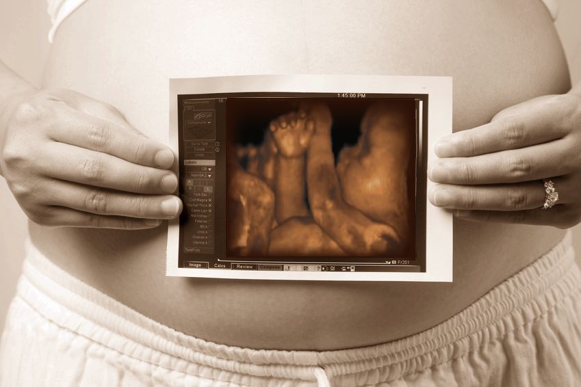
From the word itself, it gives a 3 dimensional imaging. In cases of pregnant women, a 3D ultrasound provides a 3D image of the baby growing inside of them. The 3D ultrasound offers real time imaging that is necessary for the pregnant woman as well as her baby. Thanks to this helpful equipment, significant contributions have been made in order to enhance our quality of life.
How important is a 3D ultrasound to a pregnant mother? Just around the time when a pregnant woman reaches 28 to 32 weeks gestation, she should already visit her doctor to check on the development of her baby or she could opt to visit her nearest baby center since they also have 3D ultrasounds at the ready. Remember, this technology is made available not just in hospitals; they are offered in clinics as well. The exterior of the baby is clearly visualized by the doctor as well as the mother. The 3D ultrasound is an indispensable tool in the diagnosis and management for obstetrics.
Benefits of 3D Ultrasound Did you know that it can take up to many 2D images in order to produce a 3D image? From this image, a lot can be interpreted including possible complications of pregnancy or perhaps with the development of the baby. It also is an important asset in assisting therapeutic processes since it is controlled and manipulated easily. Additionally, the device allows for more accurate visualization and provides less harmful effects than other imaging devices.
As of today, a fully interactive manipulation experience by 3D ultrasound is still in the process of development. The visualization strategies may also vary among the manufacturers. Some manufacturers offer just a single image whereas others produce multiple ones.
Before subjecting yourself to 3D Ultrasound, do take note of the basic risks of the visualization process.
Three important things to note regarding the risks of ultrasound are:
- duration
- intensity
- frequency
Generally, the first factor to take into consideration is the duration of the ultrasound exposure. Besides the fact that there is no specific time set, remember that the process does not reach for more than 30 minutes.
Although there are no reported complications to a prolonged exposure, it is best to shorten the time that the baby is exposed to the ultrasound.
The next thing to consider is the intensity of the ultrasound waves. Technically, the waves are set to a higher intensity in order to detect the heart sounds of the infant. Just like the time of exposure, the amount of ultrasound exposure should be limited as well to ensure safety.
Lastly is the frequency of sessions. It should be no more than once a month since the procedure may pose risks. Please do seek counsel from your doctor before deciding anything regarding your pregnancy and that includes the ultrasound procedure. They are definitely the ones that know what is best for you and your baby.
A Miracle Made for Pregnant Mothers – 3D Ultrasound
By Michelle Ann Reynolds
This article was brought to you by Michelle Reynolds. She brings information and solutions for those who are pregnant regarding ultrasounds and when they should or should not be performed. Find out more about 3d Ultrasounds or just visit her web site at www.ultrasoundtransducers.org.


 Make an appointment for your early gender determination or 3D/4D ultrasound today! 3D ultrasound photos and 4D ultrasound videos allow the whole family to bond with your baby!
  Be sure to purchase additional 3D ultrasound photos and 4D ultrasound video DVDs so you can share with family and friends.